Plant Cell Biology

@University of Freiburg, Germany

  • Home
  • Research
    • Intracellular infections
    • Membrane nanodomains
    • Symbiotic Remorins
    • Arabidopsis Remorins
    • Intrinsic disorder
  • Publications
  • Team
  • Service
    • Transmission Electron Microscopy
    • Correlative Microscopy
  • Teaching
    • Grundmodul (GM-01)
    • Profilmodul (PM-20)
    • Vertiefungsmodul (VM-15)
    • Wahlmodul (WM-25)
    • alle Kurse
  • Jobs
  • paper

    Yet another contribution of our EM unit

    05/02/2018

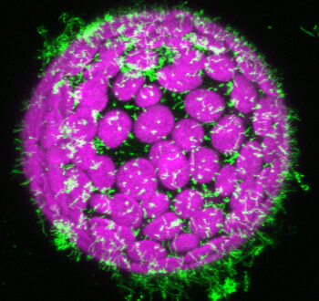
    We analyzed plastid structures from a crystal-forming Arabidopsis thaliana PHYTOENE SYNTHASE -overexpressing line by transmission electron microscopy. These roots were shown to accumulate similarly high carotenoid levels like callus. Ultrastructural analysis revealed plastid-localized membrane remnants exclusively in the carotenoid-accumulating line, while these structures were absent in WT roots. Read more in:

    Schaub P, Rodriguez-Franco M, Cazzonelli CI, Álvarez D, Wüst F, Welsch R (2018), Establishment of an Arabidopsis callus system to study the interrelations of biosynthesis, degradation and accumulation of carotenoids. PLoS ONE 13(2): e0192158.
    https://doi.org/10.1371/journal.pone.0192158

    Read More

    Related Posts

    How infection threads pass the cell wall

    08/07/2022

    New contribution of our EM lab

    13/06/2022

    New paper in Current Biology

    29/04/2021
  • paper

    New contribution of our EM unit

    23/01/2018

    In a recent publication in PNAS the group of Sonja Albers from the University of Freiburg demonstrates that the archaeal CheY protein shares the overall structure and mechanism of magnesium-dependent phosphorylation with its bacterial counterpart. Furthermore they show that phosphorylation-dependent activation, and conserved residues in the archaeal CheY helix α4, are important for interaction with the archaeal-specific adaptor protein CheF. Our EM unit contributed electron micrographs of H. volcanii archaella. Thank you Sonja for the fruitful collaboration.
    Reference:

    Quax TEF., Altegoer F, Rossi F, Lia Z, Rodriguez-Franco M, Kraus F, Bange G, and Albers SV (2018) Structure and function of the archaeal response regulator CheY.
    PNAS, doi:10.1073/pnas.1716661115

    Read More

    Related Posts

    Congratulations, Nikolaj!

    31/08/2018

    New pre-print from our lab

    29/01/2021

    Infectosome assembly by RPG during RNS

    01/03/2023
  • paper

    New findings from our lab on bioRxiv

    31/12/2017

    We have just released our newest dataset on the bioRxiv pre-print server. In this manuscript we report that the remorin SYMREM1 is required and sufficient to recruit and stabilize the Medicago truncatula entry receptor LYK3 in membrane nanodomains in a FLOTILLIN 4-dependent manner. These findings provide the molecular mechanism of SYMREM1 and demonstrate genetically that recruitment of the LYK3 receptor into membrane nanodomains is required for its function and to control host cell infection.

    Reference:
    Liang P, Stratil TF, Popp C, Marin M, Folgmann J, Mysore KS, Wen J, Ott T
    Symbiotic root infections in Medicago truncatula require remorin-mediated receptor stabilization in membrane nanodomains
    bioRxiv: doi: https://doi.org/10.1101/179036

    Read More

    Related Posts

    New contribution published in Plant, Cell & Environment

    31/05/2021

    How infection threads pass the cell wall

    08/07/2022

    New paper in Current Biology

    29/04/2021
  • paper

    Paper accepted

    10/11/2017

    We just got a manuscript accepted in Journal of Cell Science entitled “Green light for quantitative live-cell imaging in plants”. There, we together with a number of other cell biologists review advances in plant cell imaging and plant growth for microscopy studies.

    Read More

    Related Posts

    New work with contributions of our EM unit published

    13/05/2020

    New paper on proximity labelling in plants

    26/08/2020

    Congratulations, Nikolaj!

    31/08/2018
  • paper

    Paper accepted

    19/08/2017

    A comprehensive review by Thomas on “Membrane nanodomains and microdomains in plant–microbe interactions” has just been accepted for publication in Current Opinion in Plant Biology.  In this review, we not only summarize the most relevant recent findings but also suggest a novel nomenclature to precisely differentiate between types of membrane sub-compartments (i.e. membrane nanodomains and microdomains) that can be visualized in living plant cells.

    Read More

    Related Posts

    From our TEM unit: How to polarize archaea?

    10/05/2019

    New paper on proximity labelling in plants

    26/08/2020

    New contribution of our TEM unit

    12/02/2021
Newer Posts 

Recent Posts

  • May 02, 2025 Paper contribution
  • Mar 31, 2025 Innovation for medicine & plants
  • Feb 05, 2025 Welcome Henriette!
  • Jan 03, 2025 Good-bye X, see you on Bluesky
  • Nov 26, 2024 Imaging workshop
Savona Theme by Optima Themes